Here is a list of pictures taken with my Jeol JSM T100 scanning electron microscope.
All the samples have been gold coated with my plasma sputter before observation.
This instrument is so fascinating!!
Fungi spores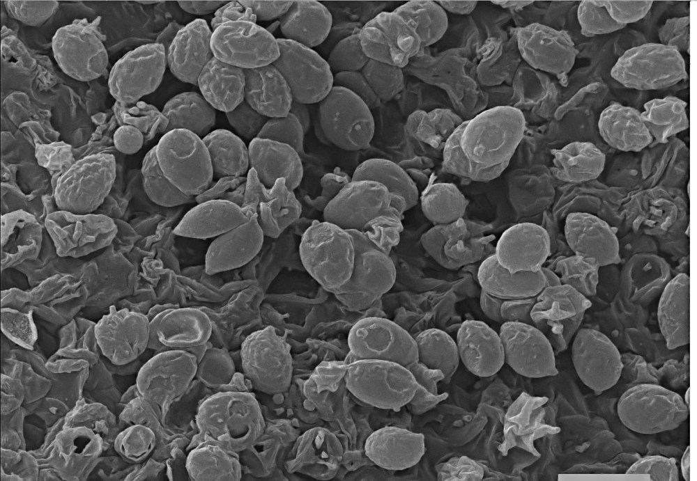
Fungi spores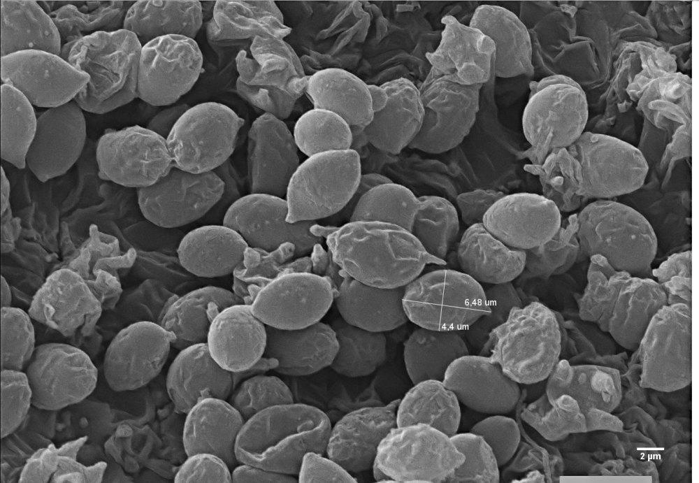
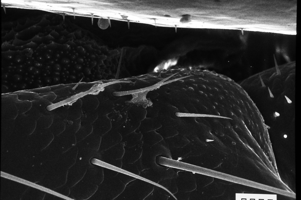
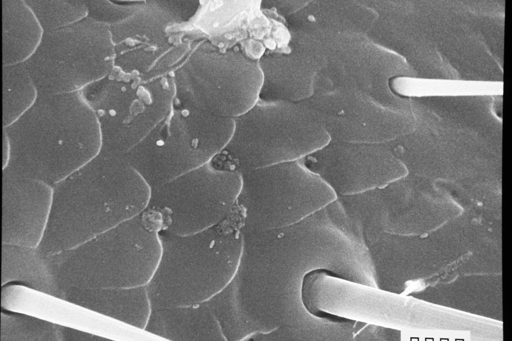
Butterfly scales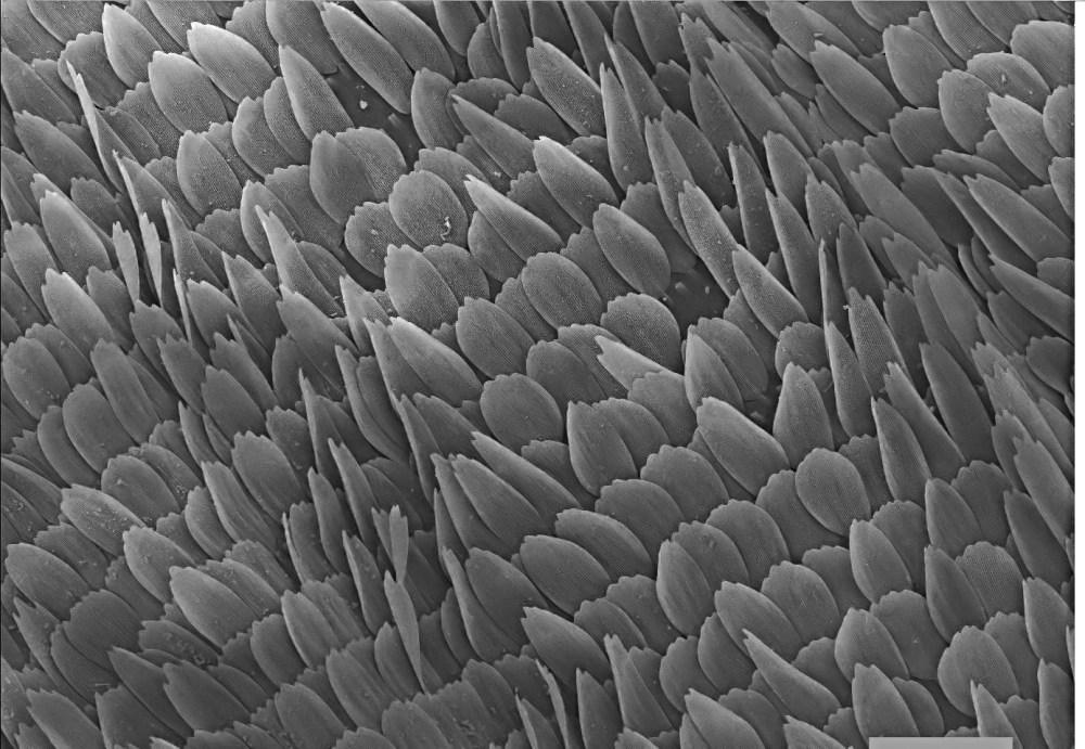
Butterfly scales, detail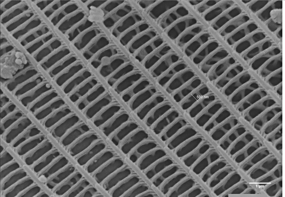
Butterfly scales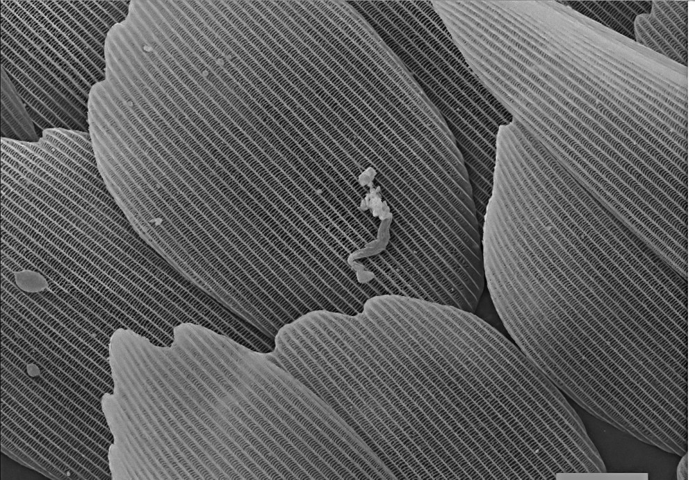
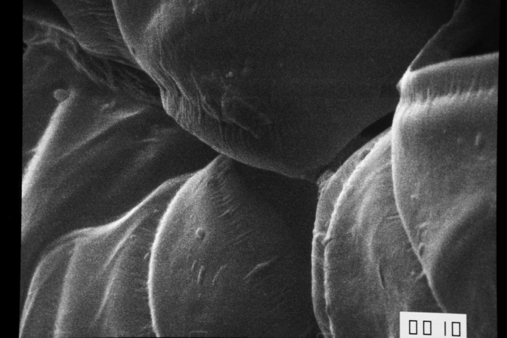
Pollen grains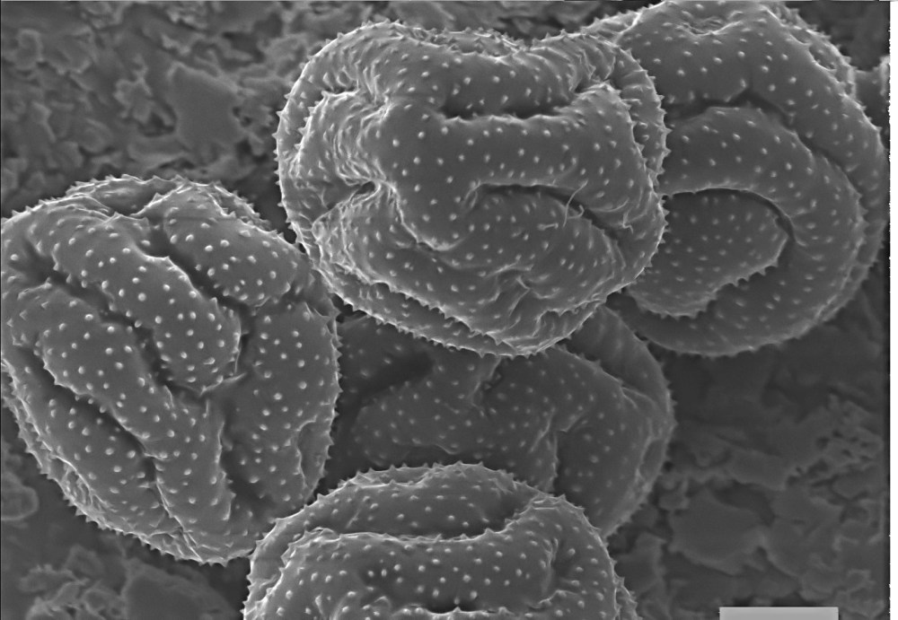
Pollen grains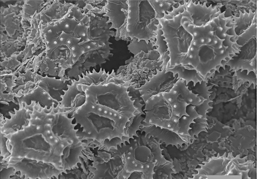
Pollen grain
Pollen grain, detail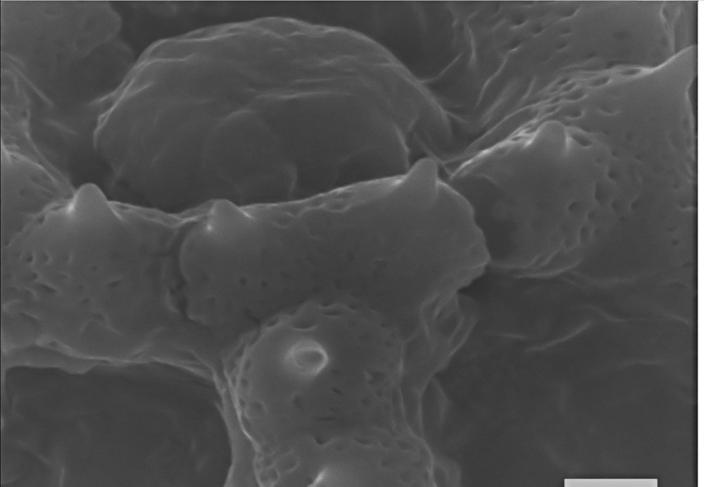
Pollen grain, detail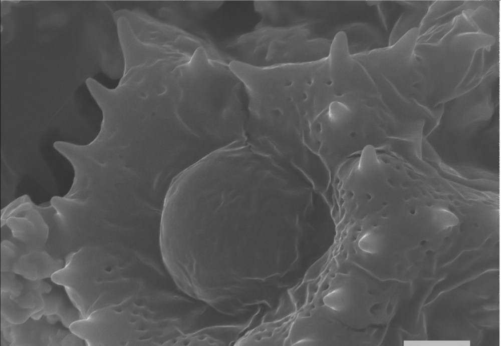
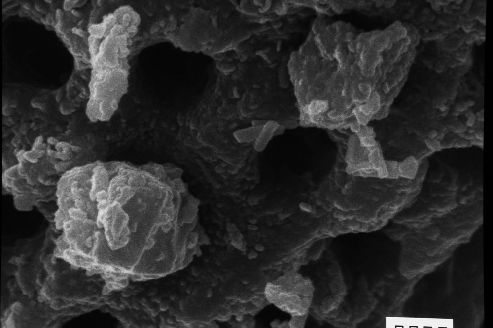
Bacteria on paper
Seashell
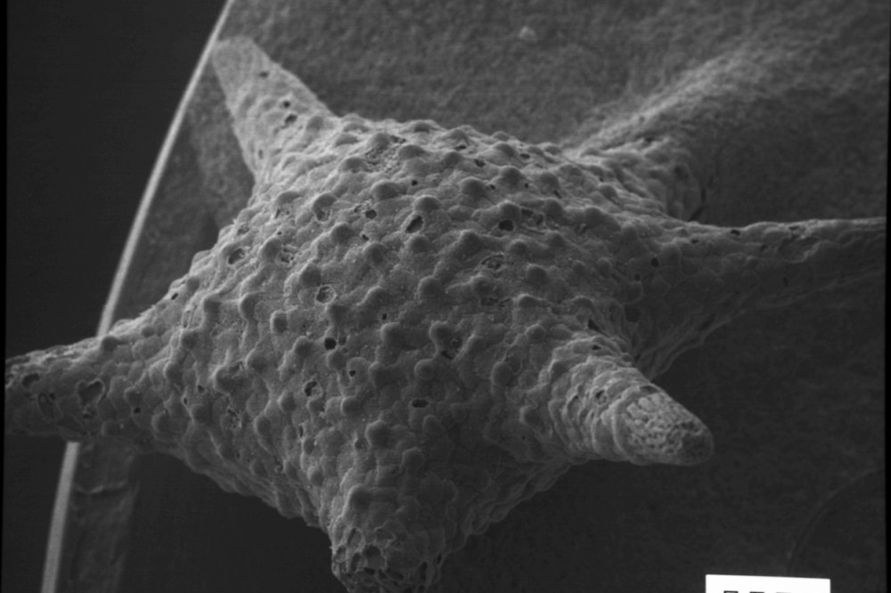
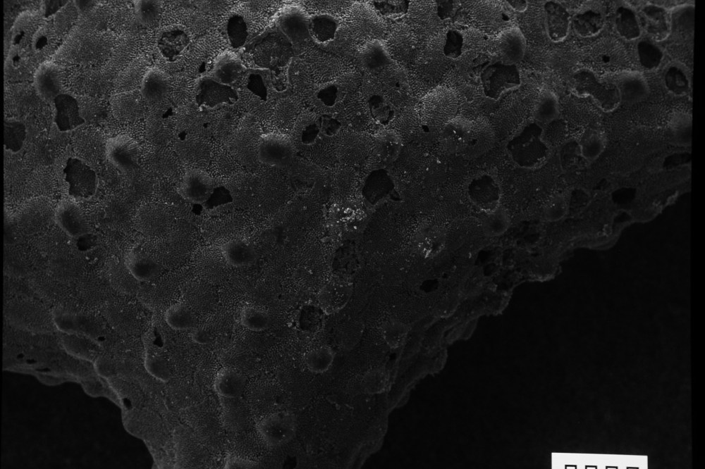
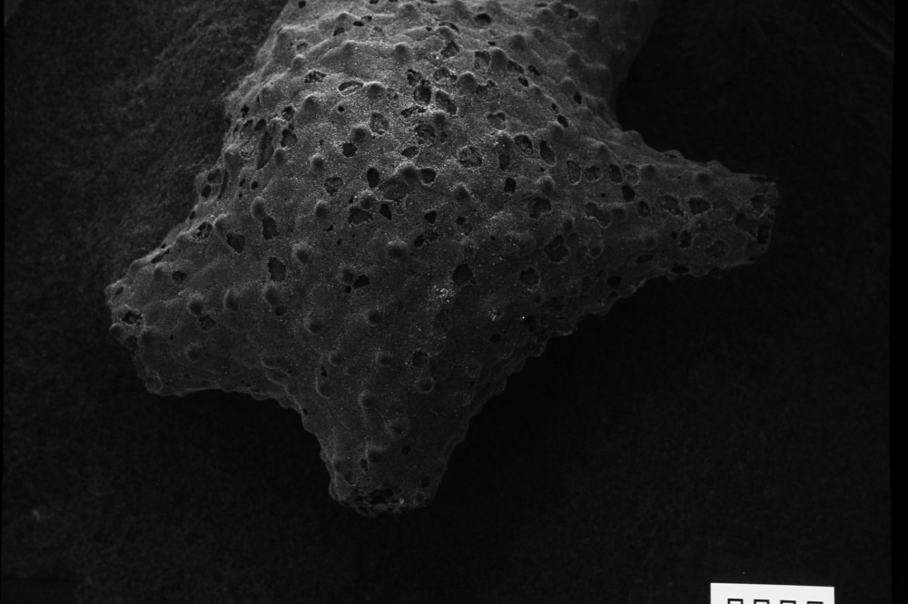
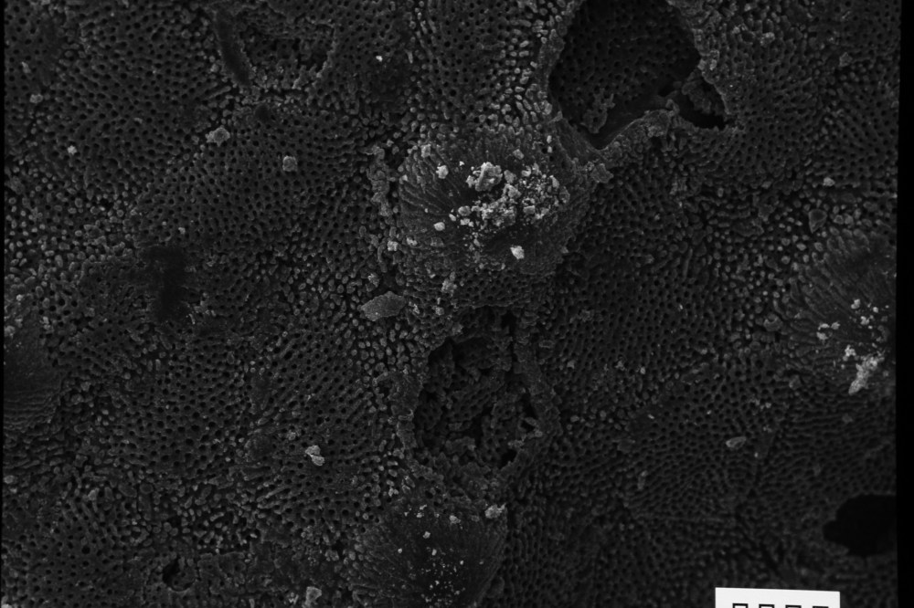
Foraminifera (Star sand, Japan)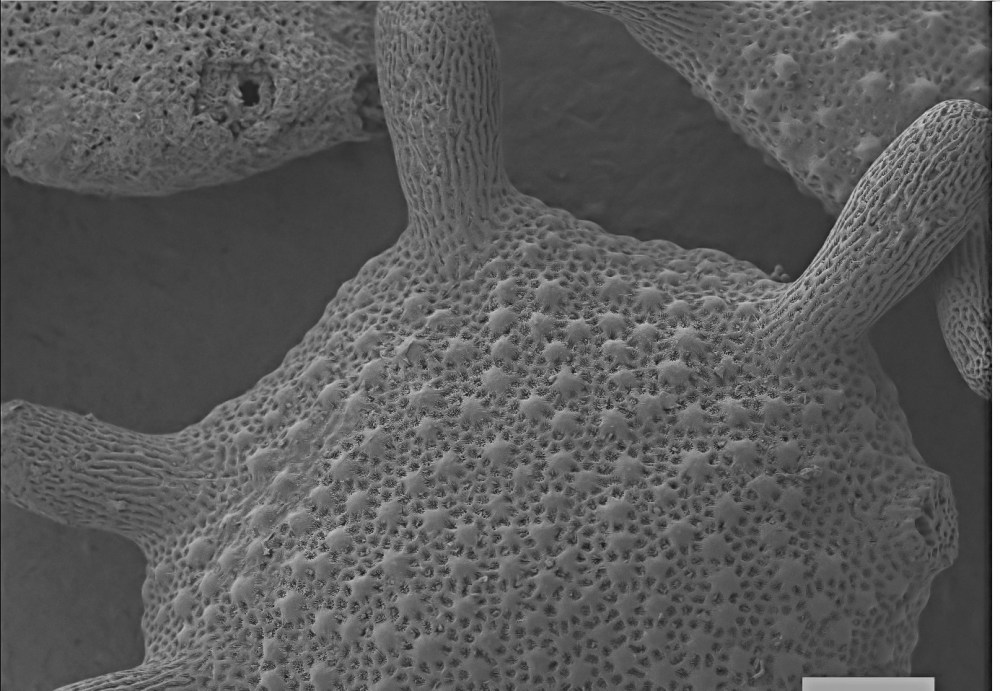
Foraminifera (Star sand, Japan), detail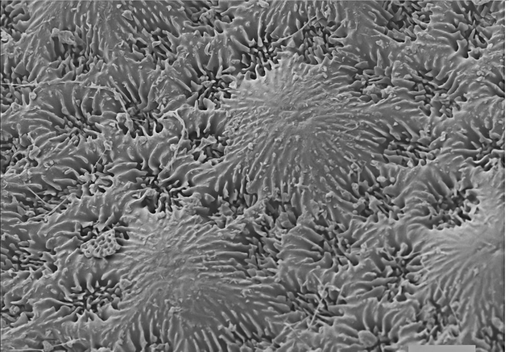
Foraminifera (Star sand, Japan), detail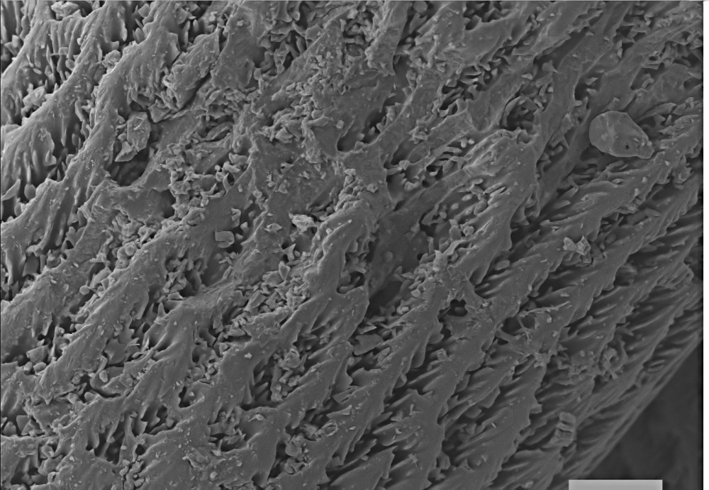
Foraminifera (Star sand, Japan), detail
Foraminifera (Star sand, Japan), detail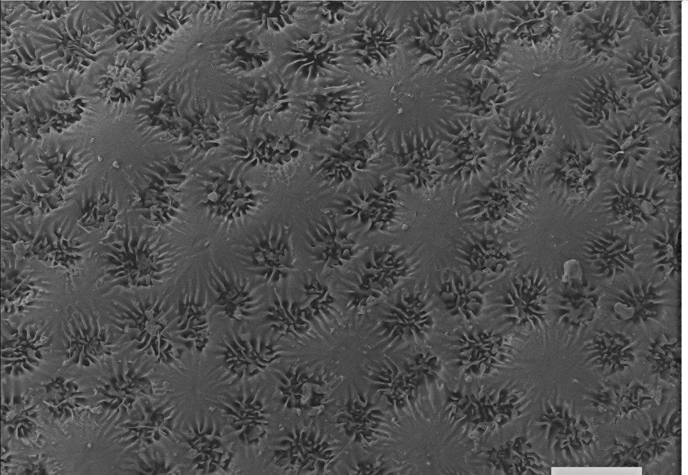
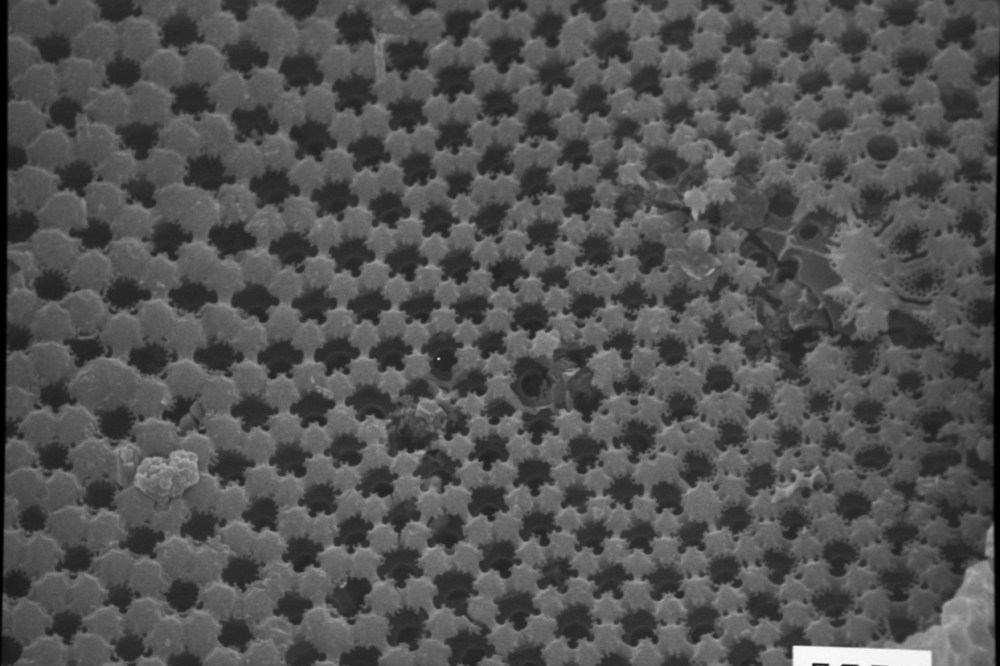
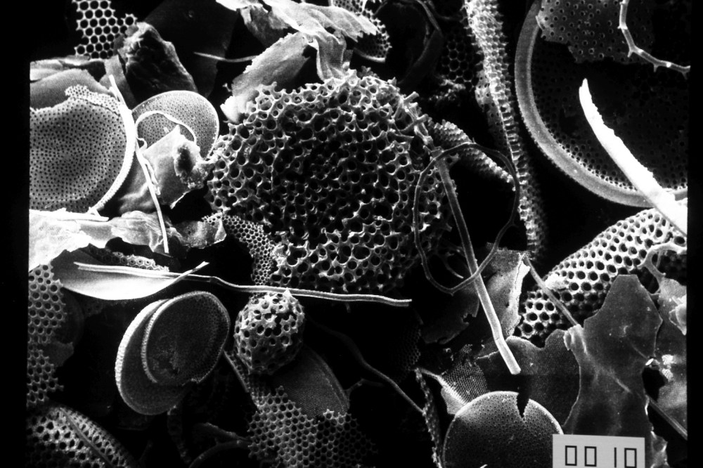
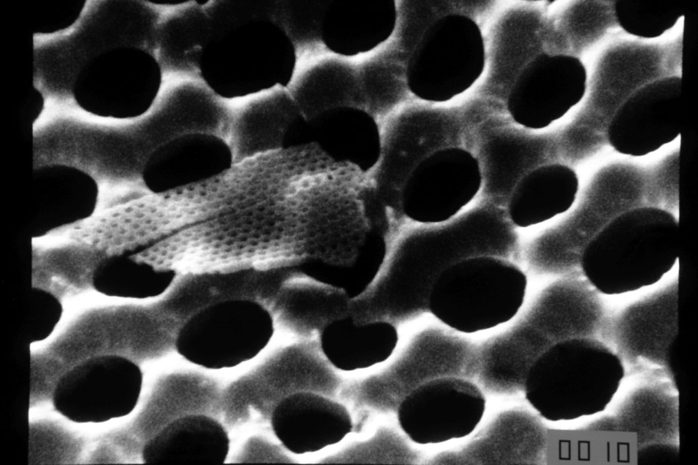
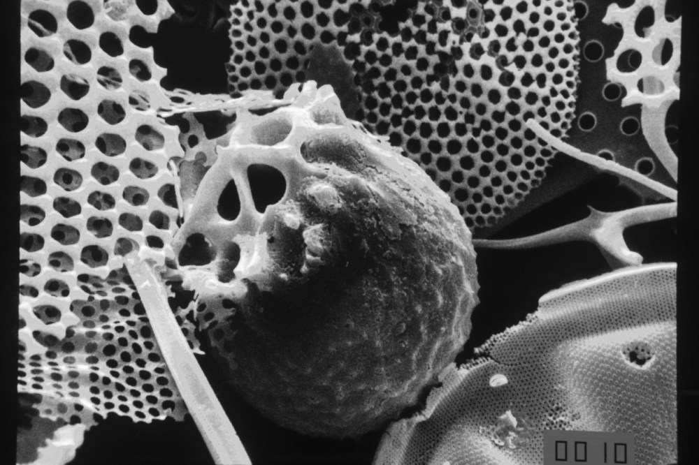
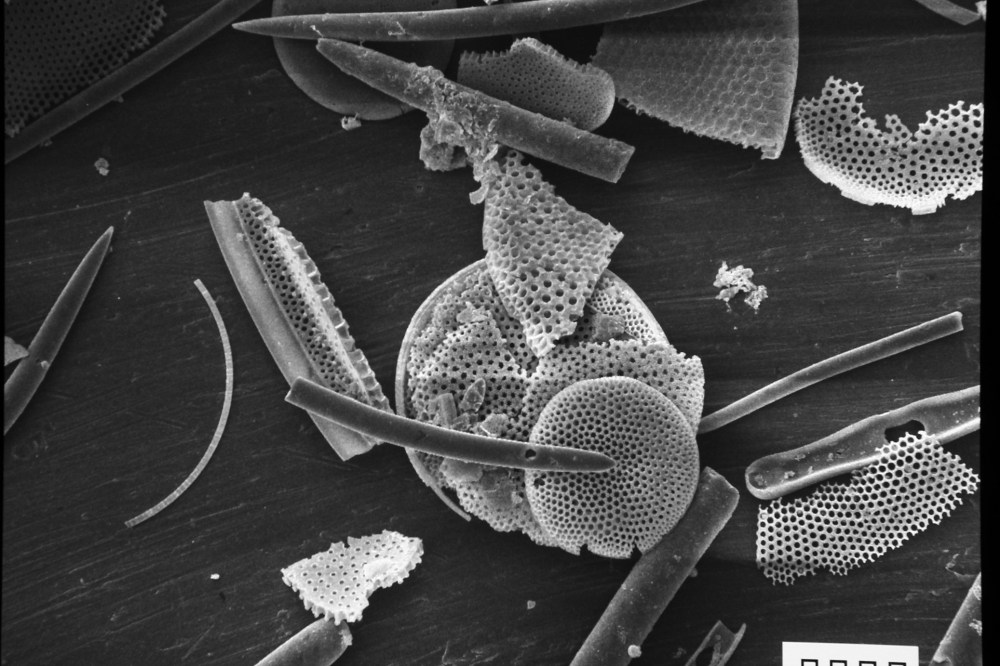
Radiolaria and diatoms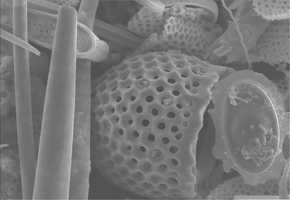
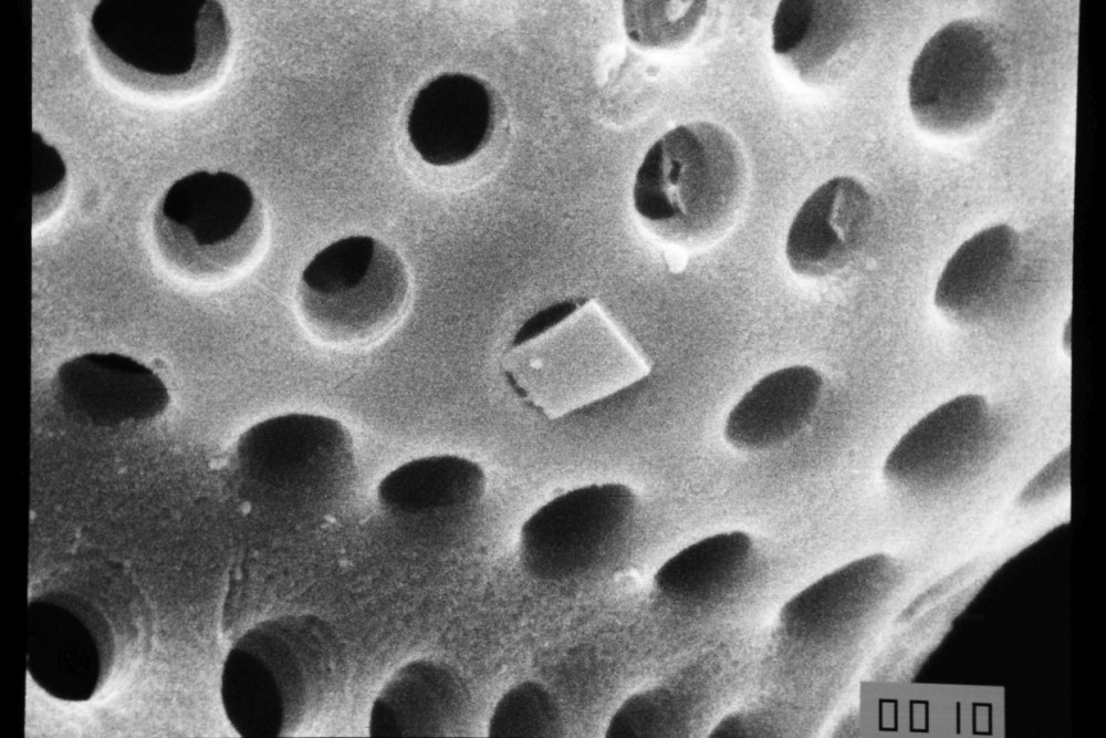
Diatom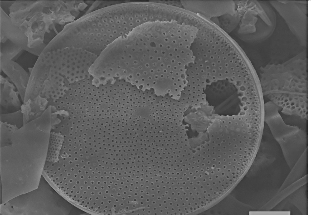
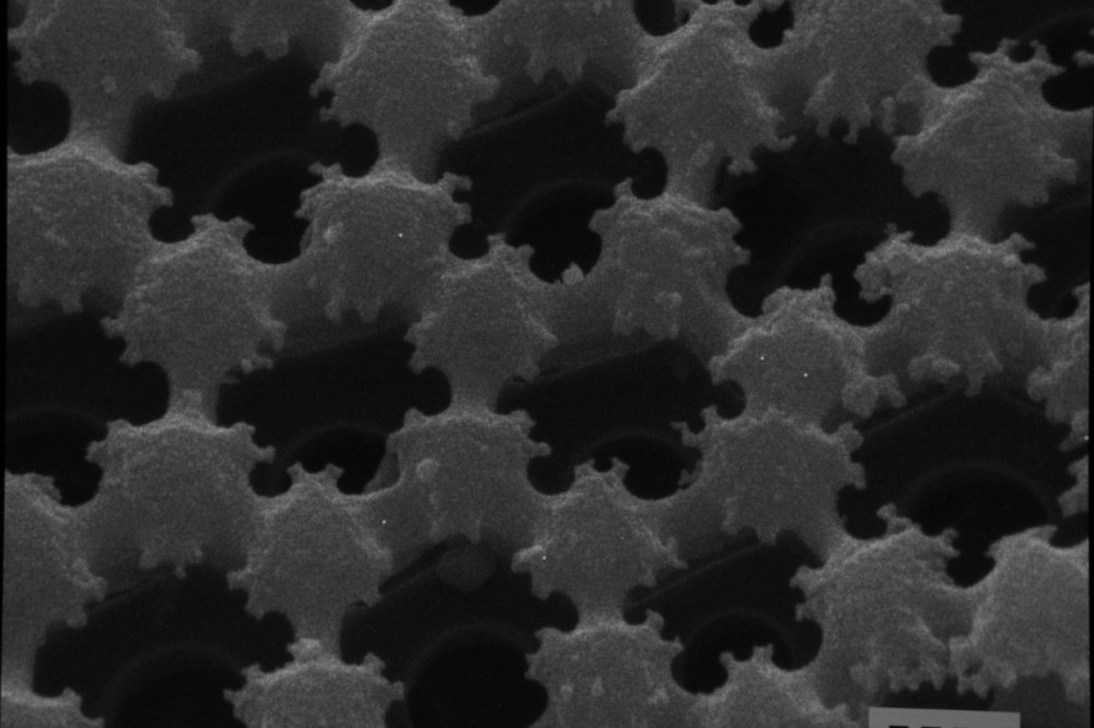
Radiolaria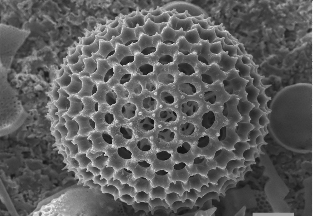
Radiolaria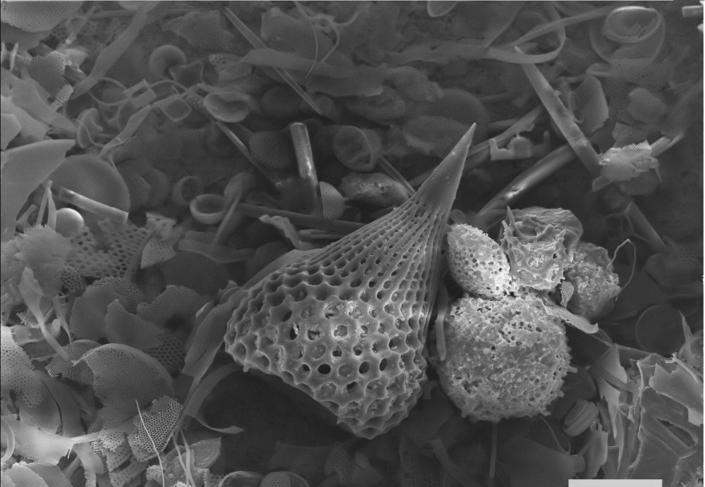
Radiolaria
Radiolaria, detail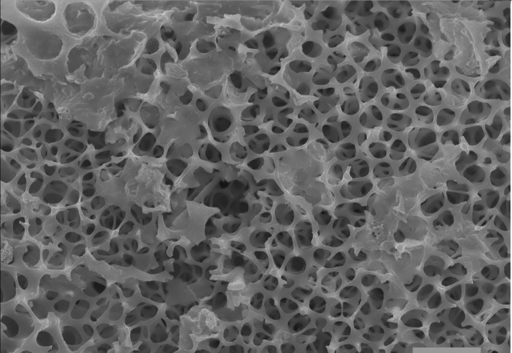
Radiolaria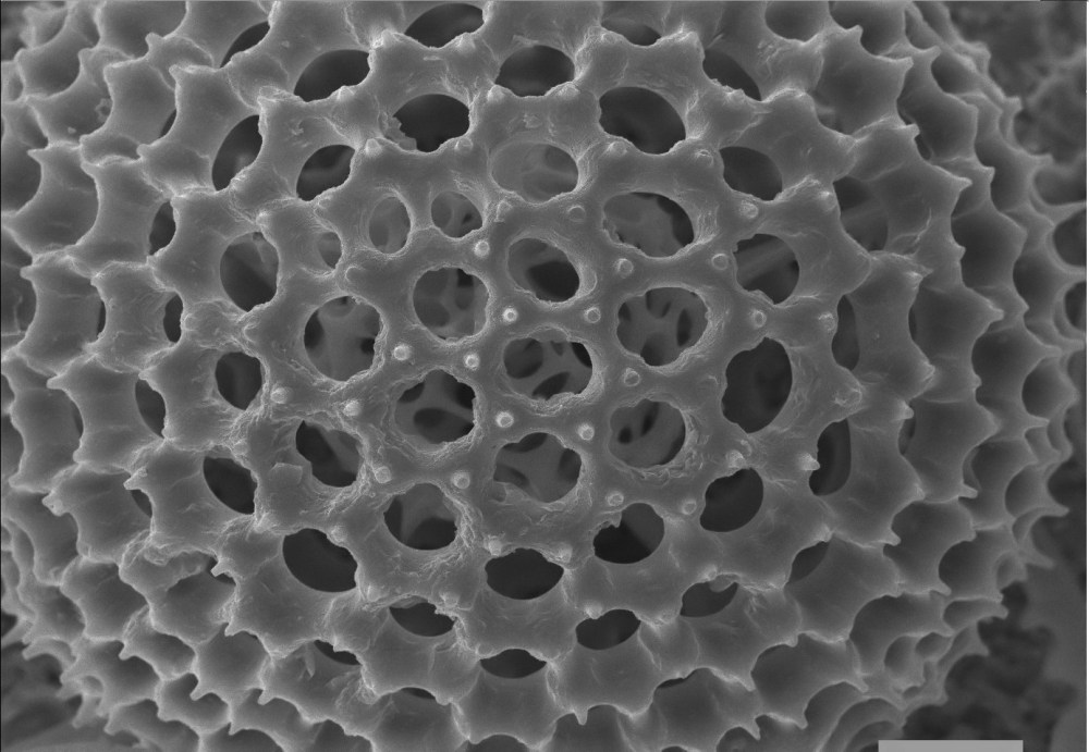
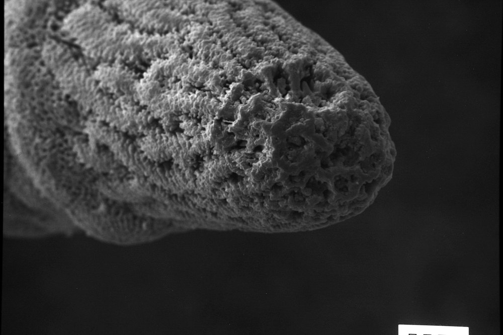
Moss sporophyte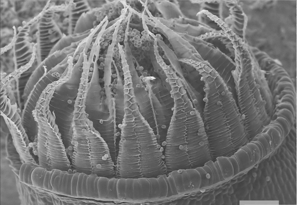
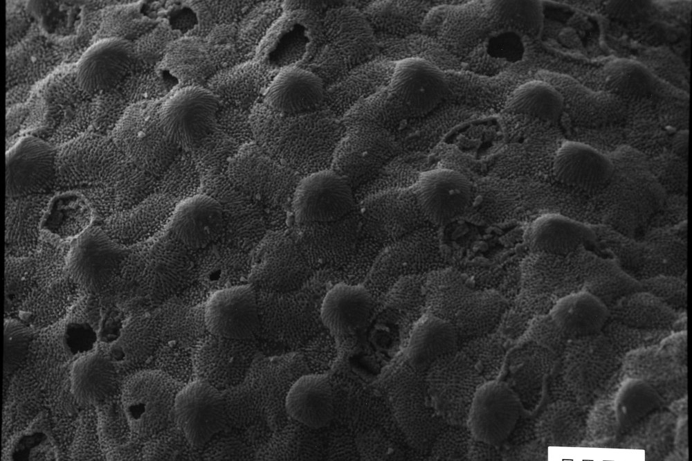
Fern spores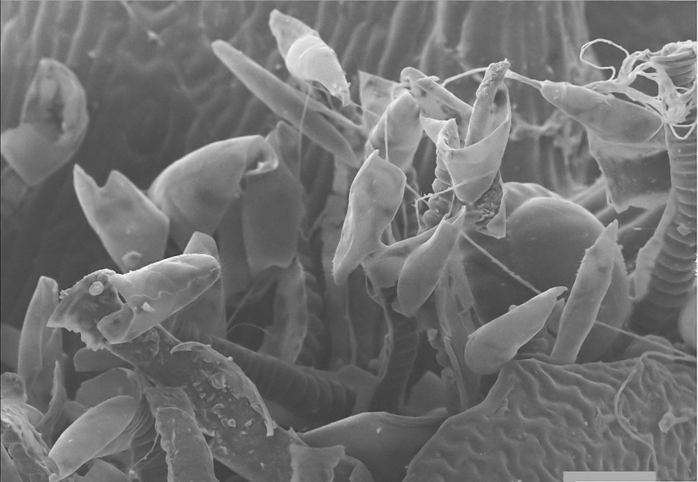
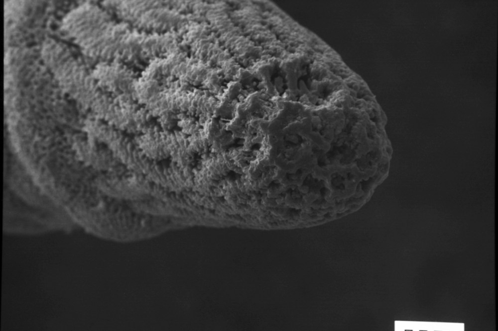
Mushroom, detail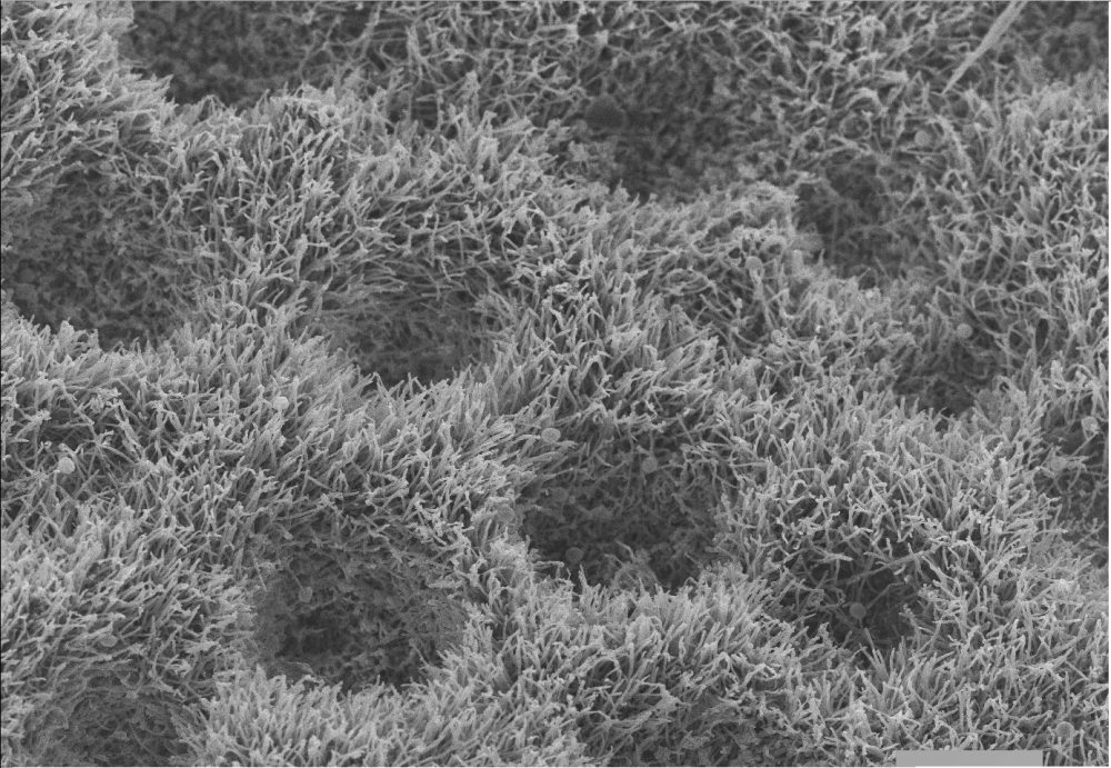
Lichen, detail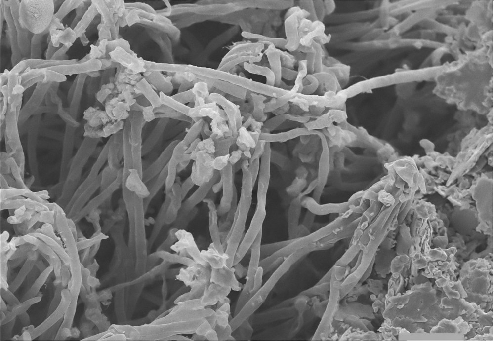
Lichen, detail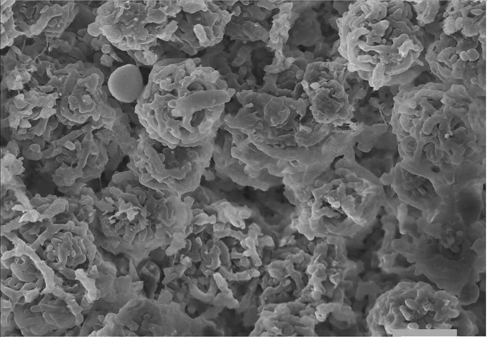
Mushroom, detail.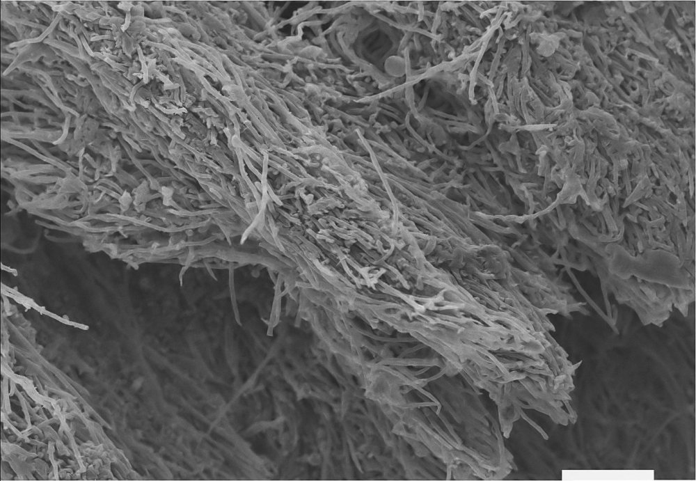
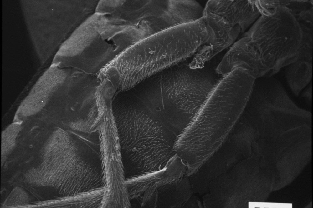
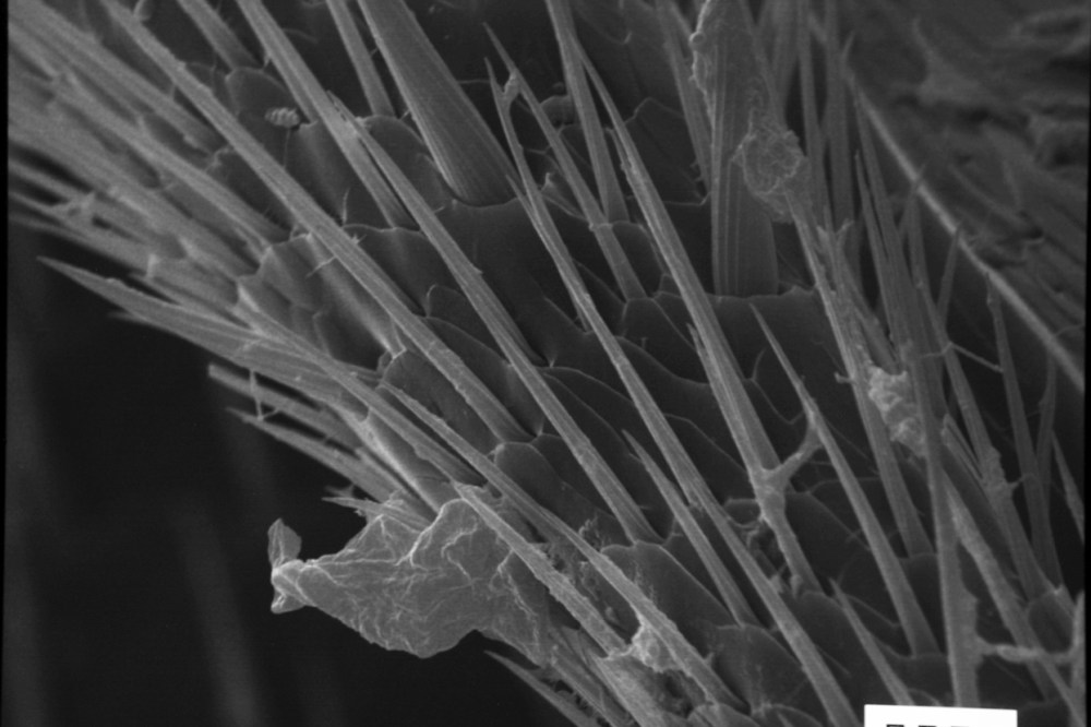
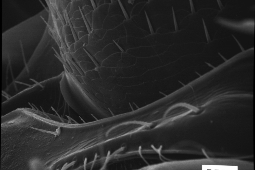
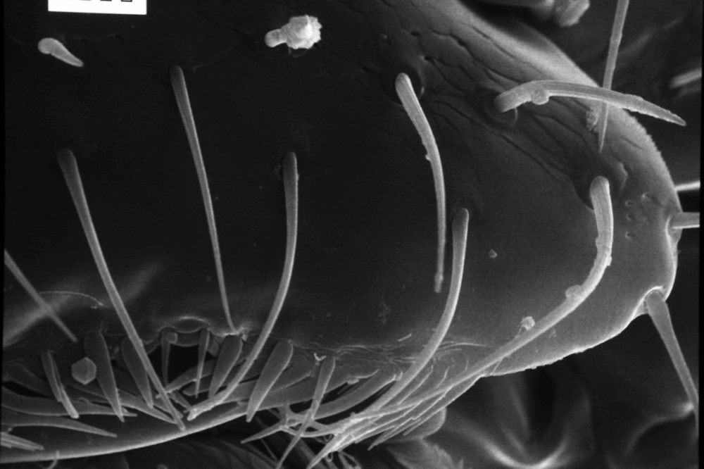
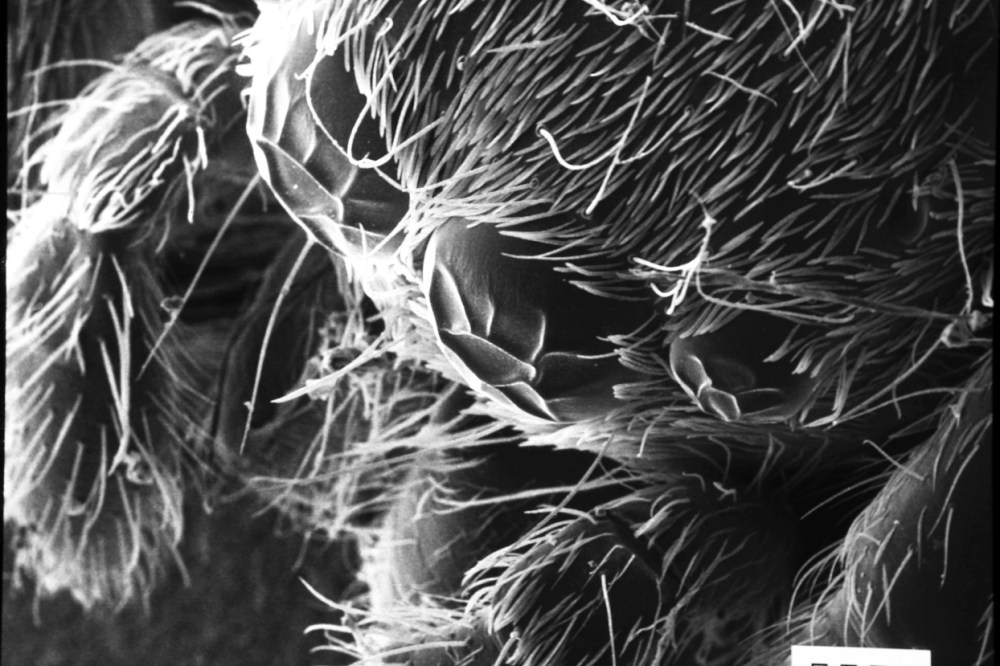
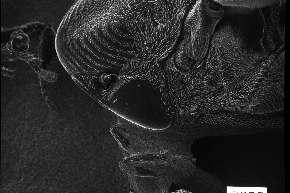
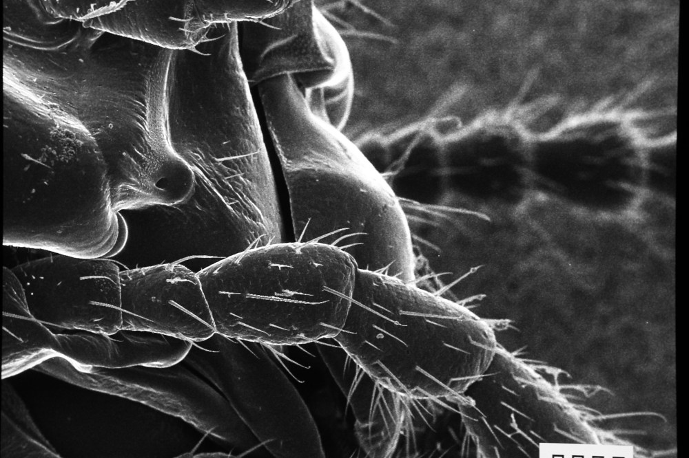
Spider head, detail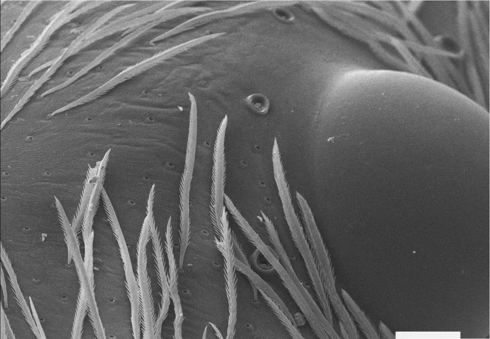
Spider head, detail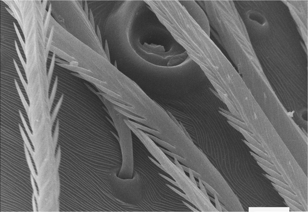
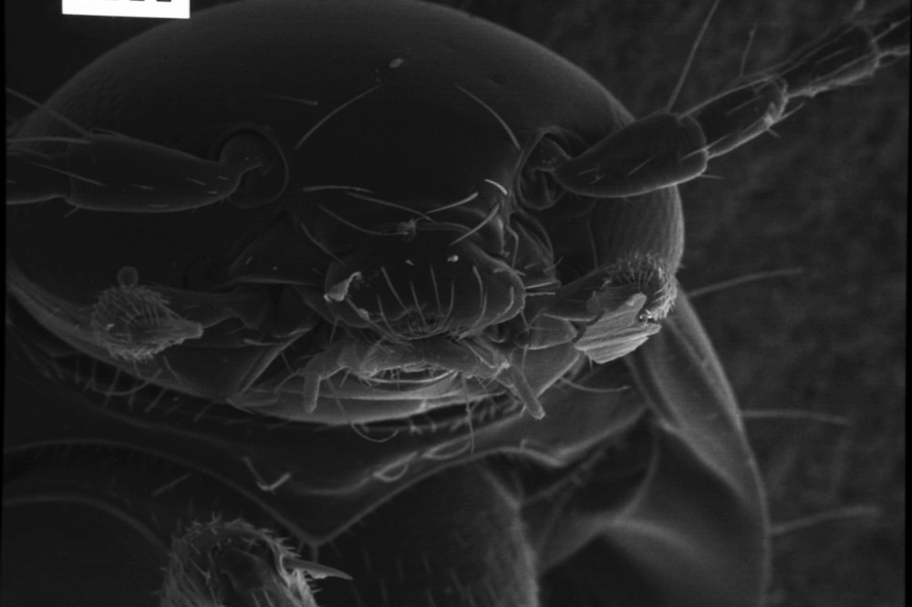
Spider head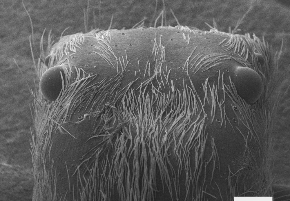
Insect head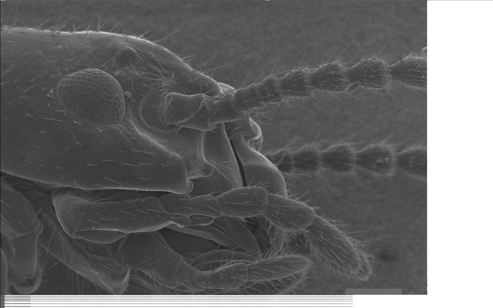
Fern stomata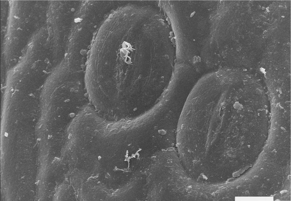
Fern stomata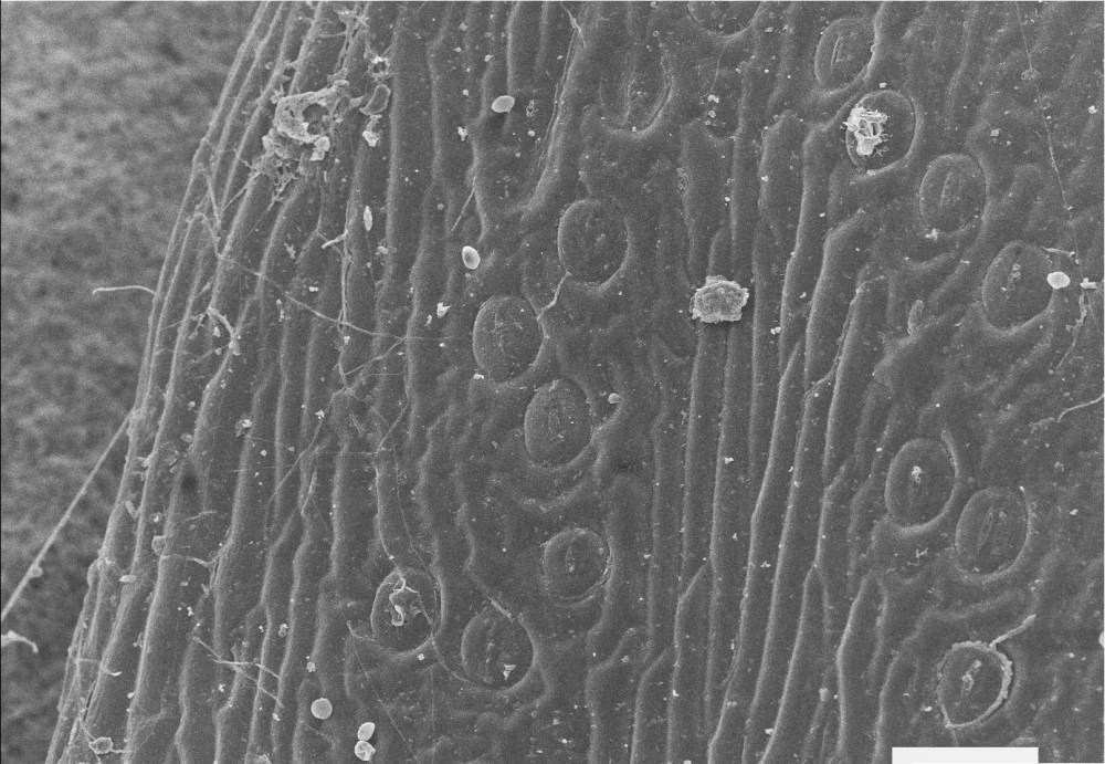
The following images have been taken with a Nikon camera syncronized with the scan of the microscope before the image digitalization upgrade.