Hello everybody! My interest in microscopy has grown up quickly since I have started to study Bryophytes in 2014. I have a bachelor’s is in Natural Sciences (University of Florence, Italy) and I have a Master in Biomedical Imaging (Turku, Finland). At the moment I’m into my PhD in Atomic and Molecular Photonics at LENS, University of Florence.
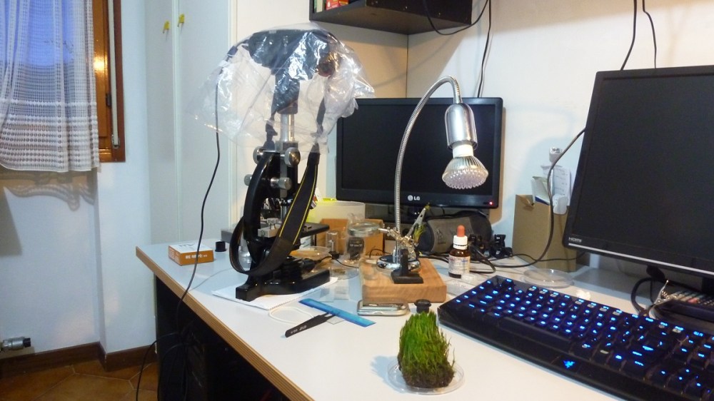
My first microscope was an old Officine Galileo, with which I took my first images of mosses. It was solid and of fine manufacture with standard RMS objectives. It has been in a box in my garage for a decade or so before I decided to remove the dust from it and find him a proper place on my working bench!


The first pictures were taken with this photo tube which allowed the use of a projection eyepiece inside it, coupled with my Nikon D300:
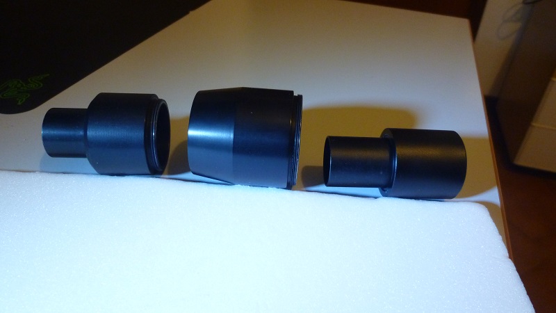


Pictures were a bit dark and with a constant light spot in the middle due to the limits of my condenser and light, but it was still possible to observe the cells and their structure. At that time I had no clue how to measure the dimensions of cells, I had no idea what ImageJ was and I was able touse Photoshop just for resizing images! 😀
After a few months I felt the need for a higher quality images and thus a new microscope was needed! I had the luck to try and buy this Hund H500 with köhler illumination, phase contrast and dark field condenser. I felt like I had the best microscope I could have ever afford! It was heavy, binocular, with a real condenser and looked so professional compared to my old Officine Galileo!


A few days after taking it home, I decided to replace the old halogen lamp with a Cree 10w Led which immediatly improved the performance of the microscope! I also had a photo tube machined which allowed the regulation in height of both eyepiece and camera.
With this scope I could take good images of my mosses and other subjects like Diatoms, resolving the striae of Amphipleura pellucida from Kemp 8 forms test.


But in those months I also started to approach the manipulation of small objects under the microscope with a very rudimental arm and needle, which it would have led me to something unexpected about one year later….

My approach to microscopes totally changed and widened after a lucky auction win of a very well preserved Zeiss Universal in August 2015.


After mastering Diatoms arrangements and common contrast techniques, I finally switched to the infinity side of the Moon with a Zeiss Axiovert 135 in 2020.

At the moment I keep arranging Diatoms and taking microphotographs of living water critters, plants, fixed specimens and many others with my instruments. Building microscopes and imaging systems as part of my PhD and my hobby.
And yes, I do own a Scanning Electron Microscope (SEM).
